Consider the cornea.
For a long time, the only way modern medicine could truly explore the structure of the cornea – the translucent, windshield-like outer part of the eye – was to cut out an entire eyeball, slice off thin layers from the front, and observe them under a microscope.
Not super practical if you wanted to examine a living patient.
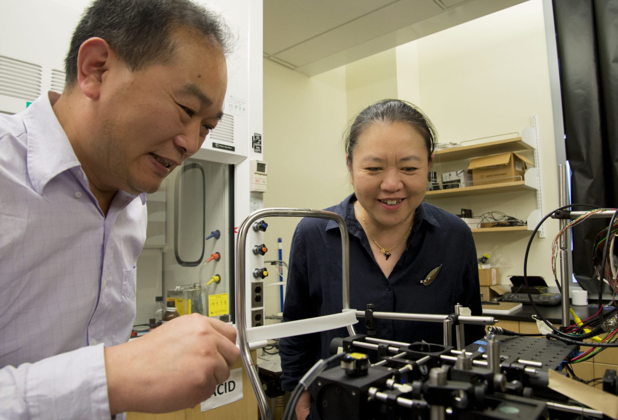
Then Ricky Wang interviewed for a job in UW’s Department of Bioengineering. Wang, a prolific inventor of biomedical imaging equipment, worked at the forefront of imaging technology that had revolutionized medicine’s ability to diagnose diseases of the retina.
During the interview, Harborview ophthalmologist Tueng Shen had a request: Can you help bring that tech to the front of the eye?
It was the start of a collaboration that’s still underway eight years later. Wang and Shen have become pioneers in unraveling mysteries from the front of the eye and developing new ways to diagnose and understand eye diseases.
“In the clinic I always say, ‘there ought to be a better way,’” Shen said. “Ricky’s expertise has helped deliver that better way.”
Their better way involves an imaging technique known as OCT, or Optical Coherence Tomography. After Wang came to UW, Shen quickly came up with a wish-list for using OCT in the clinic. Together, they’ve developed techniques and technologies to help clinicians examine patients’ corneas in unprecedented detail.
They’ve also upended conventional wisdom on how the eye works; previous models had been based on static imaging. Slices of eyeballs under a microscope gave way to dynamic 3-D visualizations of a living system.
The difference was night and day. Shen compared it to the early days of the X-ray when a chest scan would show little more than a tiny heart. Today, ultrasound and other imaging techniques let us watch the heart beat in real-time.
Speaking of heartbeats, the pair used Wang’s technology to become the first in the world to observe the flow of aqueous humor, the clear fluid between the lens and cornea. It pulsed in time with the beating of a patient’s heart.
Learning a new language
The collaboration with Shen and others across UW Medicine isn’t something Wang experienced at previous university jobs. “We produced research in a lab, looked at the results, and…that was often it,” he said. “The fun part is seeing all this work help patients in the clinic.”
To get there, though, the pair had to ditch their respective languages of engineering and medicine. Shen said she had to tone down the medical jargon. Wang didn’t write emails full of equations.
Wang admits engineers can spend their time pondering complex physics and then come back with a convoluted solution. “After working with clinicians, my view changed from thinking about high-level math to more practical solutions.”
They continue to tackle items on Shen’s wish list, which seems to keep growing. The two have an NIH grant that uses Wang’s tech to measure the strength and stiffness of corneal tissue. That could one day help identify patients who might develop complications from LASIK surgery.
Clinicians and researchers shouldn’t be shy about knocking on each other’s’ doors, Shen and Wang said. It’s fun. And it can lead to breakthroughs.
“This kind of collaboration is so important to keep us at the forefront of improving health,” Shen said.
Editor’s note: Shen and Wang discussed their collaboration at an event celebrating the 50th anniversary of the Department of Bioengineering.